Roof Of Lesser Sciatic Foramen
What Are The Bony Structures Th And More Terms From B3 Anatomy Flashcards Memorang

Anat Gluteal Posterior Thigh Flashcards Quizlet

Greater Sciatic Foramen Google Search Medical Terminology Study Lower Limb Technology Life

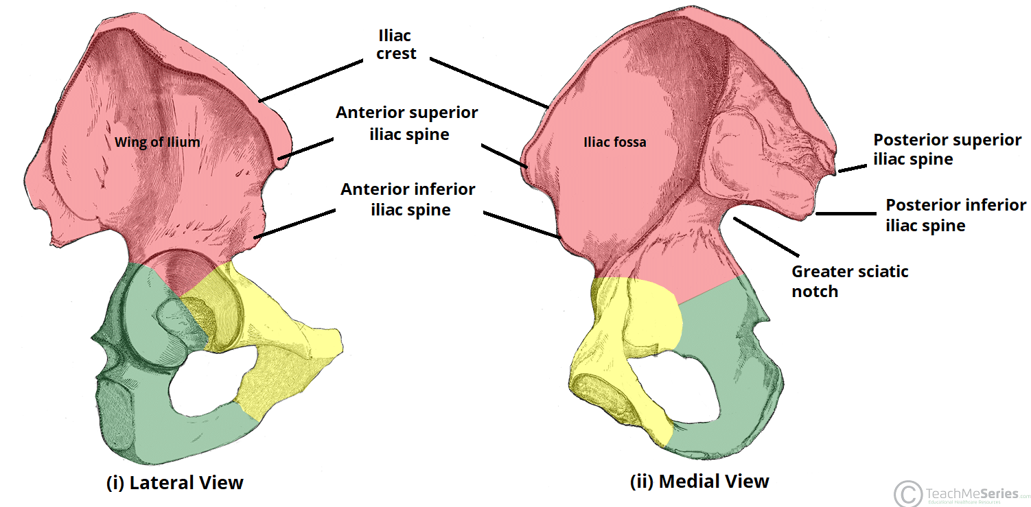
Bony Framework Of Pelvis Anatomy Sacral Promontory Transverse Processes Of Lumbar Vertebrae Iliac Tuberosity I Pelvis Anatomy Anatomy Bones Anatomy Images

Sciatic Foramen Piriformis Lower Limb Muscle

Unit 36 Gluteal Region Post Thigh And Popliteal Fossa At Saint Louis University Studyblue
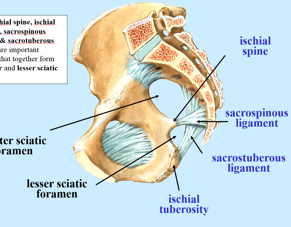
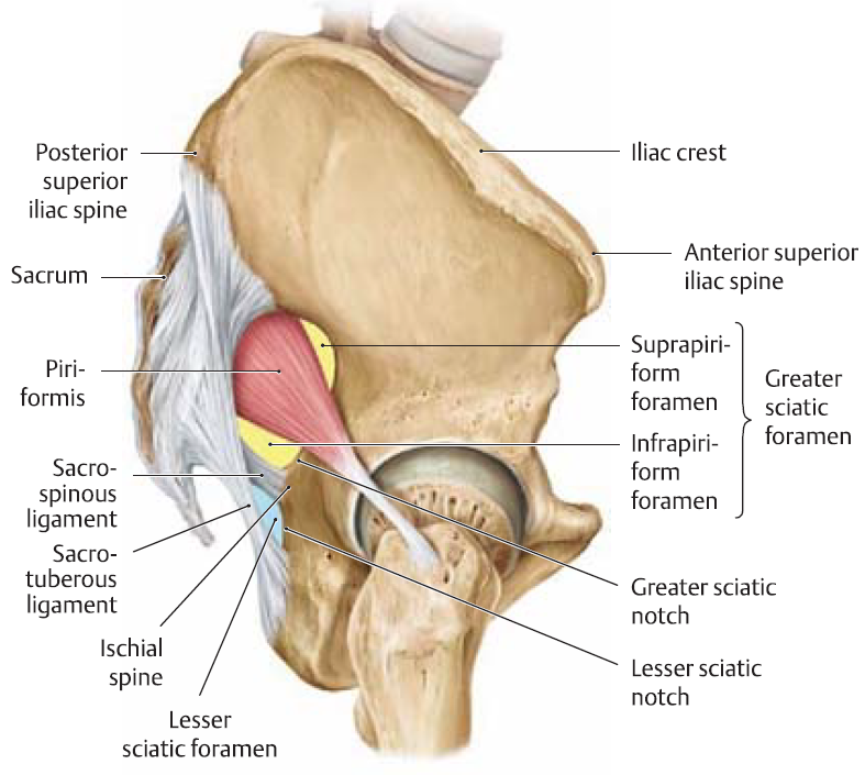
The sacrospinous ligament separates the greater sciatic and lesser sciatic foramina.
Roof of lesser sciatic foramen.

Pudendal Nerve Note Behind Sacrospinous Ligament In Relation With Ischial Tuberosity Alcocks Canal Note Pudendal Nerve Passes Through Both Greate
Archie S Lower Limb Regions And Drawings Flashcards Memorang

Anatomy Of The Pudendal Nerve Health Organization For Pudendal Education Pelvis Anatomy Pelvic Bone Anatomy Bones

Gastrulation Dote Anatomy Topics
Source : pinterest.com
